
In Austria, complex tumour operations are planned using a 3D model in virtual reality (VR). The software for this was developed at the University of Basel.
Doctors must not get too close to a tumour: "If we cut into the tumour, it is absolutely fatal for the patient," says Michael Nogler, Deputy Director and Head of the Department of Experimental Orthopaedics at Innsbruck University Hospital. The surrounding tissue, for example nerves, must also not be injured. If an incision is too deep, the patient may no longer be able to move their leg or hold back their urine after the operation.
In order to take these factors into account before the procedure, conventional two-dimensional X-ray computed tomography (CT) and magnetic resonance imaging (MRI) images of the patient are used. While these are normally analysed on screen, VR now enables a much more precise examination.
3D model with software from Basel
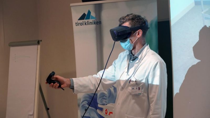
Doctors at the Innsbruck University Clinic for Orthopaedics recently prepared the first operation in Austria in virtual space. The operating doctor puts on VR goggles and can use them to analyse the entire anatomy of the tumour patient in three dimensions. The new visualisation makes it easier to calculate how the tumour can be removed from the body completely and without major consequences.
The way the software developed at the University of Basel works is not fundamentally new: as is usually the case, the CT and MRI data is superimposed in layers to create volume models or anatomical representations. The software is particularly fast at rendering this data. The 3D visualisation can then be inspected from all sides. "What is new is the way in which it is brought into virtual space, creating a new user interface that we can work with collaboratively," says Nogler.
This means that not only those wearing VR glasses can enter the room - the recording can be transmitted to a screen for all those present. According to the specialist, international experts can also be connected live during preparation. The analysis is carried out ad hoc and without manual post-processing, as is often required for conventional analyses of layered image data.
Zoom possible
The main advantage is that the doctor can plan the procedure under almost real conditions and see the patient's body exactly as it will be during the operation: in 3D. "Viewing three-dimensional information in virtual space is simply much more intuitive," says the expert. The visualisation can also be rotated and flipped using a joystick - the doctor can also walk around the display and zoom in on the tumour, for example.
The very first operation was painstakingly prepared several months in advance and ultimately performed successfully. During the risky procedure, a young patient had a football-sized bone tumour removed from his hip and lumbar vertebrae. Since then, the method has been used regularly at Innsbruck University Hospital.
The future
According to Nogler, the next step is to fully integrate the innovation into the university hospital's X-ray system. In the future, it is also conceivable that the entire operation could be played out in detail in advance: "We can't yet document and simulate all the steps in the software," says Nogler. They are currently being carried out mentally.
Source: Futurezone