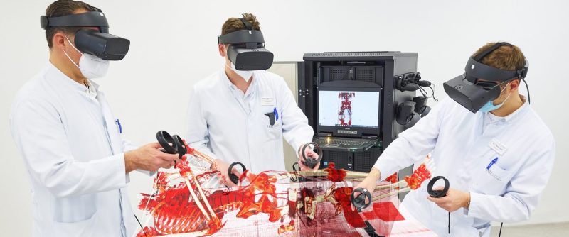
Two assistant doctors from the Department of Surgery at the University Hospital Bonn (UKB) have developed a tool with which medical students can now experience surgical teaching content in virtual reality (VR) space. Equipped with VR glasses, students can take on the surgeon's view of a patient and thus explore anatomy in the virtual 3D world. Surgical treatment concepts thus become understandable in a simple and intuitive way.
Four medical students and a resident stand at the front of the lecture hall, equipped with VR headsets on their heads. They all look around a bit uncoordinated, turn from time to time and reach into the air around them with their hands. Dr Jan Arensmeyer and Philipp Feodorovici from the Department of General, Visceral, Thoracic and Vascular Surgery at the UKB have been holding this very special seminar since the summer semester of 2021.
VR in constant use
The two assistant doctors specialising in thoracic surgery have been working on it together with senior physician PD Dr Philipp Lingohr and senior physician Dr Nils Sommer from the surgical clinic's teaching team since they received funding from the state of NRW for the virtual teaching pilot project. With the support of Prof. Joachim Schmidt, Head of Thoracic Surgery at the UKB, and in close cooperation with the Zurich software company MedicalholodeckSince the first use of the VR tool last semester, it has been constantly adjusted and improved. In the meantime, the VR technology is reliably in use almost every Wednesday in the lecture theatre of the surgery department and is continuously being further developed despite limited resources.
Understanding of anatomy and disease patterns
Complex 2D representations from imaging procedures - such as MRI or CT - can be experienced three-dimensionally in the virtual world. The students learn the anatomy of the skeleton, the vessels or the internal organs artificially processed in three dimensions, until then mainly from the textbook. In the advanced stage of studies, the focus is then on the interpretation of imaging diagnostics (X-ray, CT, MRI) and presents great challenges, especially for the inexperienced viewer. "We can now close this gap. By viewing the imaging reconstructed in three dimensions in real time in the VR-Space, and thus simulating the surgeon's detailed view of a patient's body, the students at the University of Bonn can develop a good understanding of the real anatomy and the various clinical pictures and thus learn the surgical treatment approach much more quickly," says Dr Arensmeyer.
And the fun factor is not neglected either when using VR goggles teaching. The students need little training time and learn to handle images intuitively in a relaxed atmosphere in the VR Space. According to initial evaluations, well over 90 percent of the students who have participated so far would like to see work with VR glasses permanently integrated into everyday clinical practice and teaching.
In the future from afar
In the near future, it should also be possible to experience virtual interventions risk-free via the tool and to meet virtually with users from other locations. "At the moment, the VR units are still location-bound, but our vision for the future is that we will also be able to offer participation in VR lessons from any location, e.g. from home. Experts or lecturers from different clinics could also meet in the virtual world to discuss a patient's case and plan the operation, with the students participating as observers. Remote learning', says Feodorovici, can thus also gain a high status in medical education."
Source: idw-online


